Beyond the Nobel Prize: Shining a light on the protein that revolutionised brain research
The 2026 Nobel Prize in Medicine celebrates discoveries that have made it possible to study brain activity using light. By introducing the light-sensitive protein Channelrhodopsin-2 into nerve cells, researchers could use light to control the cells’ activity, giving rise to the field of optogenetics. But what actually happens to the protein itself when the light is switched on?
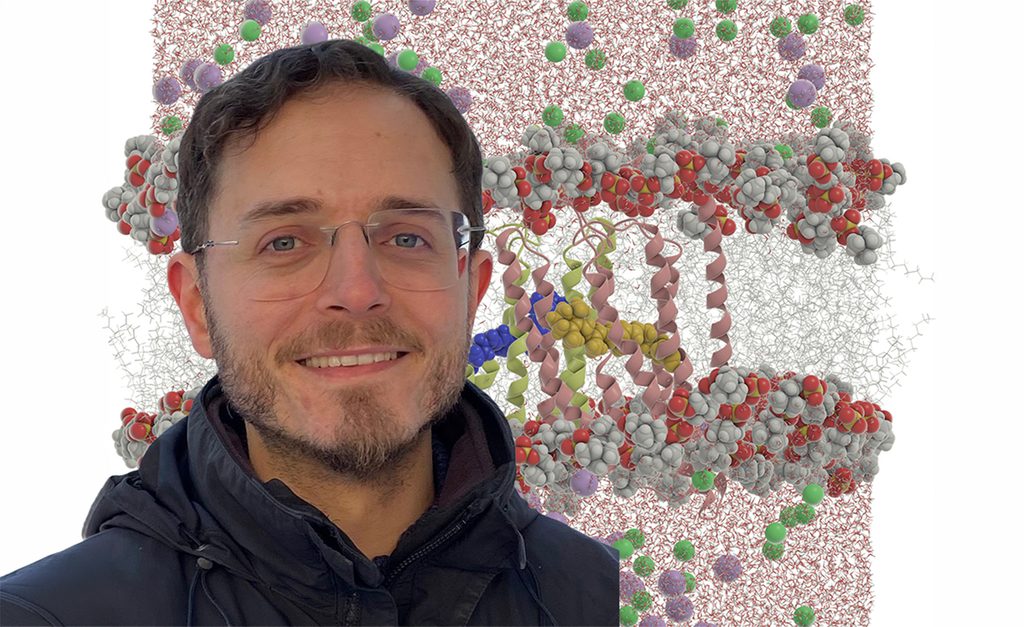
For David Carrasco de Busturia, a theoretical chemist and Marie Skłodowska-Curie Postdoctoral Fellow at KTH Royal Institute of Technology, the Nobel Prize has brought new attention to the questions that already drive his research. Just two weeks before the Nobel Prize announcement, he presented his work at a conference in Siena, where he got the chance to discuss Channelrhodopsin-2 with Peter Hegemann – the Nobel laureate who co-discovered the protein.
“So it was especially exciting to hear the news two weeks later,” says Carrasco de Busturia. Understanding the protein in molecular detail, however, is a considerable computational challenge. Using resources provided by NAISS, he is carrying out large-scale simulations to investigate exactly how Channelrhodopsin-2 responds to light.
A light-sensitive biological switch
Channelrhodopsins are light-sensitive proteins originally found in microorganisms such as algae. Embedded in the cell membrane, they act as tiny channels controlling the flow of ions into the cell. At the centre of the protein is retinal, a molecule also found in our own eyes. When retinal absorbs light, it changes shape, triggering the protein channel to open and altering the activity of the cell.
This mechanism became the basis for optogenetics, where light-sensitive proteins are introduced into cells to control their activity using light. But being able to use Channelrhodopsin-2 is not the same as fully understanding how it works.
“We understand the protein well enough to use it as a tool in optogenetics, but we still don’t completely understand all the steps it goes through, from that first light to the opening and eventual closing of the channel,” he says.
A quarter of a million atoms
The challenge is partly one of scale. Whilst the retinal molecule itself consists of only 63 atoms, it sits inside the much larger Channelrhodopsin-2 protein, which in turn is embedded in a membrane, surrounded by water and ions. Altogether, his computational model comprises around a quarter of a million atoms.
To add to that, the processes span dramatically different timescales. When retinal absorbs light, the molecule changes shape within 100 femtoseconds, while subsequent changes in the protein unfold over nanoseconds and eventually microseconds. “Capturing that range of timescales is part of what makes simulating the process so challenging,” he says.
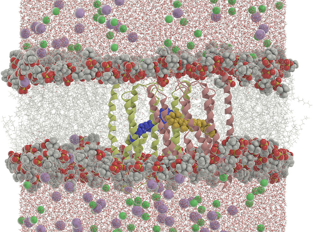
In his simulations he uses a technique known as QM/MM. This allows him to use quantum mechanics (QM) for the parts of the system where quantum effects are crucial, like the changing retinal molecule, while describing the much larger surrounding protein, membrane and water environment with less computationally demanding molecular mechanics (MM) models.
In this way, he can zoom in with very high physical detail where that detail matters most, without having to perform the same expensive quantum calculation for every atom in the system.
Where NAISS comes in
Even with this division of labour, simulating the full molecular system requires considerable computing power. Carrasco de Busturia uses Dardel, the NAISS computing resource hosted at KTH, where the computational work is divided across many processor cores running in parallel.
“For scientific software development or even for setting up the protein in its lipid membrane environment, I can use my own laptop, but to simulate the steps the system goes through when light hits, I really need a supercomputer,” he says.

Although being fundamental research, knowledge generated today about the molecular mechanisms of rhodopsins, he points out, could help guide the future development of better, more efficient optogenetic tools, improving our understanding of the brain and neurodegenerative disorders.
“One of my main goals is to model and simulate the whole movement of the system and the photocycle it goes through,” he says, likening it to producing a molecular-scale movie of Channelrhodopsin-2 in action.
Reaching that point will require not only powerful computing infrastructure, but increasingly efficient software capable of using that computing power. For now, each simulation brings him another step closer to seeing how one of biology’s remarkable light-sensitive machines works.
Read more specifics on David Carrasco de Busturia’s NAISS project.
Read more user stories.
David Carrasco de Busturia acknowledges funding from the European Union’s Horizon Europe research and innovation program under the Marie Skłodowska-Curie grant agreement No. 101199012.